Enlargement of axillary accessory breast tissue is during pregnancy and lactation. The presence of galactocele in the axillary accessory breast is a rare occurrence 4 5.

Sonographic Findings Of Accessory Breast Tissue In Axilla And Related Diseases Lim 2017 Journal Of Ultrasound In Medicine Wiley Online Library
Accessory breast tissue itself is normal and should not be misdiagnosed as an abnormality.

. Accessory breast tissue results from failed regression of primitive mammary tissue and is most often located in the axilla. Accessory breast tissue derives from a failure of the primitive mammary tissue to regress after development of the mammary ridge in the thoracic area at week 7 of gestation. Ectopic breast tissue is at risk for benign and malignant breast disease with reported diagnoses of fibrocystic disease mastitis fibroadenoma atypical hyperplasia and carcinoma 46.
When found on mammography accessory axillary breast tissue should be recognized as a normal developmental variant rather than considered a pathologic lesion although carcinoma can develop in the accessory tissue. 4 9 10 and can be appreciated on mammography ultrasound and MRI. These lesions include soft tissue masses associated with nontumorous conditions accessory breast tissue and chronic granulomatous.
Furthermore the palpable lump showed indeterminate characteristics U3 measuring 109 83 mm Fig. The most common reason for seeking surgical. The axillary accessory breast tissue develops as part of polymastia along the milk line.
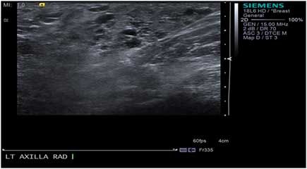
After childbirth AAB engorgement and hyperplasia may occur due to milk formation. Figure 2 shows the normal sonographic appearance of accessory breast tissue in the axilla. Both benign and malignant diseases that occur in the normal breast can also develop in accessory breast tissue in the axilla.
Most women are unaware of their accessory breast tissue and it is detected incidentally on a mammogram. Both benign and malignant diseases that occur in the normal breast can also develop in accessory breast tissue in the axilla. It is found anywhere along the milk lines with axillary localization being most frequently reported.
A female presented with a history of the right axillary region pain and swelling during a premenstrual time. Radiographically the accessory tissue resembled the remaining normal glandular tissue but was separate from it. Accessory breast tissue itself is normal and should not be misdiagnosed as an abnormality.
This is of common occurrence in women. Accessory Breast Tissue on Ultrasound. Accessory breasts consist of ectopic breast tissue that results from a failure of the embryonic mammary ridge to regress.
The diagnosis of axillary masses can be challenging because various tumors can develop in parts of the axilla other than lymph nodes even though we frequently encounter axillary masses in daily practice. We evaluated the optimal time for AAB excision and assessed variations in clinical. Symptoms associated with an axillary accessory breast AAB may newly develop or worsen after pregnancy.
The combined survival beyond the 4-year post-treatment period was 94. 6 rows Failure of involution of the milk line results in accessory breast tissue most commonly in. 1 The variability of presentation and the possibility of other disease make this problem clinically challenging and although it is a well-known entity there is no established classification system to guide its management.
A total of 90 cases of carcinoma of ectopic breast tissue were found 64 of which occurred in the axilla. Up to 10 cash back Background Accessory breasts are usually located in the axilla. Sonographic image of a palpable area of concern in the left axilla demonstrates normal dense fibroglandular tissue consistent with prominent accessory breast tissue.
Discomfort pain milk secretion axillary thickening and local skin irritation can occur. They are most common in the anterior axillary line 1 2 and are subject to the same disease processes as normal breast tissue. Axillary breast tissue affects between 2 and 6 percent of women.
Accessory breast tissue occurs in 26 of women Fig. The ultrasound shows accessory breast tissue in the axillary without any associated pathology. Failure of involution of the milk line results in accessory breast tissue most commonly in the axilla but it can occur anywhere from the axilla to the inguinal region.
Accessory breast tissue responds to hormonal stimulation and may become more evident during menarche pregnancy or lact. Accessory breast AB is defined as the presence of extra and ectopic breast tissue. Ultrasound scan of the right axilla showed accessory breast tissue measuring 56 50 mm in its maximum dimension without any abnormal axillary lymph nodes.
Accessory breast tissue results from failed regression of primitive mammary tissue and is most often located in the axilla. Accessory breasts are classically distributed along the embryonic milk line. Although it is most commonly located in the axilla ectopic or accessory breast tissue may be seen anywhere along the thoracoabdominal milk line.
Up to 10 cash back Accessory axillary breast tissues are common embriogenic alterations found in 1 to 6 of women that often manifest bilaterally 12. The clinical presentation can be from asymptomatic to cyclical changes. AB is thought to be an embryonic mammary tissue remnant that can occur with or without the nipple and areola 23.
2 Determining which surgical technique to use is critical to achieving an optimal. The appearance of accessory breast tissue on ultrasound irrespective of location is the same as the appearance of breast tissue within the breast. Accessory breast tissue itself is normal and should not be misdiagnosed as an abnormality.
At least 6 of the population has accessory breast tissue in. Accessory breast tissue results from failed regression of primitive mammary tissue and is most often located in the axilla. When accessory tissue occurs both benign and malignant breast lesions.
Axillary accessory breast usually presents as bilateral swellings in the axilla. The mean radiographic dimension of the accessory tissue which was best seen on oblique or. Both benign and malignant diseases that occur in the normal breast can also develop in accessory breast tissue in the axilla.
A specific radiography-aided diagnosis of accessory axillary breast tissue can eliminate unnecessary biopsy. Mammographic features of normal accessory axillary breast tissue were analyzed in 13 women 54 of whom had positive findings on physical examination. This line extends from the axillary region down to the groin.

Sonographic Findings Of Accessory Breast Tissue In Axilla And Related Diseases Lim 2017 Journal Of Ultrasound In Medicine Wiley Online Library

Accessory Breast Tissue Radiology Reference Article Radiopaedia Org
Accessory Axillary Breast Tissue

Sonographic Findings Of Accessory Breast Tissue In Axilla And Related Diseases Lim 2017 Journal Of Ultrasound In Medicine Wiley Online Library
Ultrasound Scan Of Right Axillary Accessory Breast Tissue Shows Focal Download Scientific Diagram

Accessory Axillary Breasts And Supernumerary Nipple Radiology Case Radiopaedia Org

Accessory Breast Tissue Radiology Reference Article Radiopaedia Org

Sonographic Findings Of Accessory Breast Tissue In Axilla And Related Diseases Lim 2017 Journal Of Ultrasound In Medicine Wiley Online Library
0 comments
Post a Comment